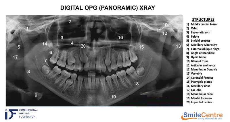
An OPG (also knows as Orthopantomagram or Panoramic radiography or panoramic X-ray or Panorex) is a two-dimensional (2-D) Panoramic Scanning Dental X-ray examination that captures the entire mouth in a single image, including the teeth, upper and lower jaws, jaw joints, and surrounding structures and tissues. This is often conducted before the commencement of dental treatment for several reasons:
- Comprehensive View: An OPG X-ray provides a wide and detailed view of the entire upper and lower jaws, including the teeth, jawbone, and temporomandibular joints. It captures a panoramic image, allowing dentists to assess comprehensive dental and skeletal structures.
- Diagnosis of Dental Issues: OPG X-rays assist in diagnosing various dental conditions, such as dental caries (cavities), periodontal disease (gum disease), impacted teeth, abscesses, cysts, tumours, and fractures. The image reveals information that may not be visible during a regular dental examination, enabling dentists to make accurate diagnoses.
- Treatment Planning: OPG X-rays aid in treatment planning by providing dentists with essential information about the position, alignment, and condition of teeth, as well as the relationship between the upper and lower jaws. This information is particularly valuable for procedures like orthodontic treatment, dental implants, extractions, and oral surgery.
- Evaluation of Bone Density: OPG X-rays can assess the density and quality of the jawbone. This is crucial when considering dental implant placement or other procedures that require a stable bone structure for successful outcomes.
- Monitoring Dental Development: In cases involving children or adolescents, OPG X-rays help monitor the development of permanent teeth and eruption patterns and identify potential orthodontic issues if any. This allows for timely intervention and appropriate treatment planning.
- Screening for Pathologies: OPG X-rays can aid in detecting pathologies, such as cysts, tumours, and abnormalities in the jaw or surrounding structures. Early identification of these conditions is vital for timely referral to specialists and appropriate management.
- Baseline imaging of the maxillofacial region: By obtaining this baseline OPG X-ray, dentists can establish a reference point for future comparisons. It helps them track changes in the teeth and jaws over time, monitor the progression of dental conditions, and plan appropriate treatment strategies. It aids in the diagnosis, treatment planning and monitoring of dental health.
Limitations of OPG X-ray
While digital OPG X-rays offer numerous advantages over traditional film-based X-rays, they accomplish some limitations. Here are a few limitations of digital OPG X-rays:
- Image Resolution: Digital OPG X-rays may have a lower resolution when compared to conventional intraoral X-rays. Minor particulars, such as small cavities or subtle changes in bone density, may be less visible in digital OPG images. Intraoral X-rays taken with film or digital sensors placed inside the mouth, can provide higher resolution for close-up views of specific areas.
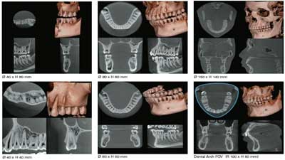
- Limited Diagnostic Information: Digital OPG X-rays provide a panoramic overview of the entire jaw, but may not provide detailed information about specific teeth or individual roots. Intraoral X-rays or other imaging techniques like cone-beam computed tomography (CBCT) are often needed to obtain more precise information about localized dental conditions.
- It is two-dimensional: The digital OPG X-ray being a two-dimensional X-ray will not help in providing a three-dimensional orientation as well as imaging of structures located at various levels or depths. For three-dimensional and detailed imaging of structures, CBCT (cone beam CT) imaging will have to be carried out.