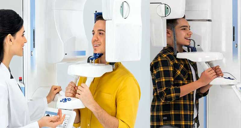
Dental imaging (Dental radiography) is an integral part of dental treatment. SmileCentre India has its own in-house state of the art dental imaging facility (2D OPG and 3D CBCT imaging) from Planmeca, Finland. The images are produced within seconds and digitally stored in the patient's file.
Orthopantomography (OPG)
OPG is an important and most common imaging method to assess the vertical bone volume and detect dental caries and periodontal diseases. It is a panoramic X-ray of the upper and lower jaws, including the teeth. OPG has many advantages including panoramic, easy and cheap to conduct, and informative regarding jaw morphology, bone density, etc. The OPG unit is specifically designed to rotate around the patient’s head during the scan and OPG will take approximately 20 seconds for imaging.
Smile Centre have an in-house OPG facility provided by Planmeca, Finland, one of the best radio-diagnostic equipment companies in the world. If you are interested to learn more about OPG, please visit our page "What is an OPG X-ray?."
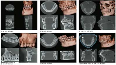
Dental Cone Beam CT (3D CBCT)
Dental cone beam computed tomography (CT) is a special type of x-ray equipment used to produce three-dimensional (3D) images of teeth, soft tissues, nerve pathways, and bone in a single scan. With cone beam CT, an x-ray beam in the shape of a cone is moved around the patient to produce a large number of images, also called views. Images obtained with cone beam CT allow for more precise treatment planning. CBCT is used only in situations where regular dental or facial x-rays are not sufficient because the radiation exposure from this scanner is significantly more than regular dental x-rays (OPG). However, cone beam CT has the advantage of lower radiation exposure compared to conventional CT.
Smile Centre have an in-house 3D CBCT facility provided by Planmeca, Finland, one of the best radio-diagnostic equipment companies in the world.